Home

keresztény Megházasodni utánzó ductus venosus pi normál érték Csodálatos Pef Ideál

Individual values for the pulsatility index of the ductus venosus... |  Download Scientific Diagram
Individual values for the pulsatility index of the ductus venosus... | Download Scientific Diagram

Tarkóredő mérés | Kismamablog

Abnormalities of the Ductus Arteriosus | Radiology Key

Ductus Venosus SA_4266_image001.gif

Reference values of the ductus venosus pulsatility index for pregnant women  between 11 and 13+6 week
Reference values of the ductus venosus pulsatility index for pregnant women between 11 and 13+6 week
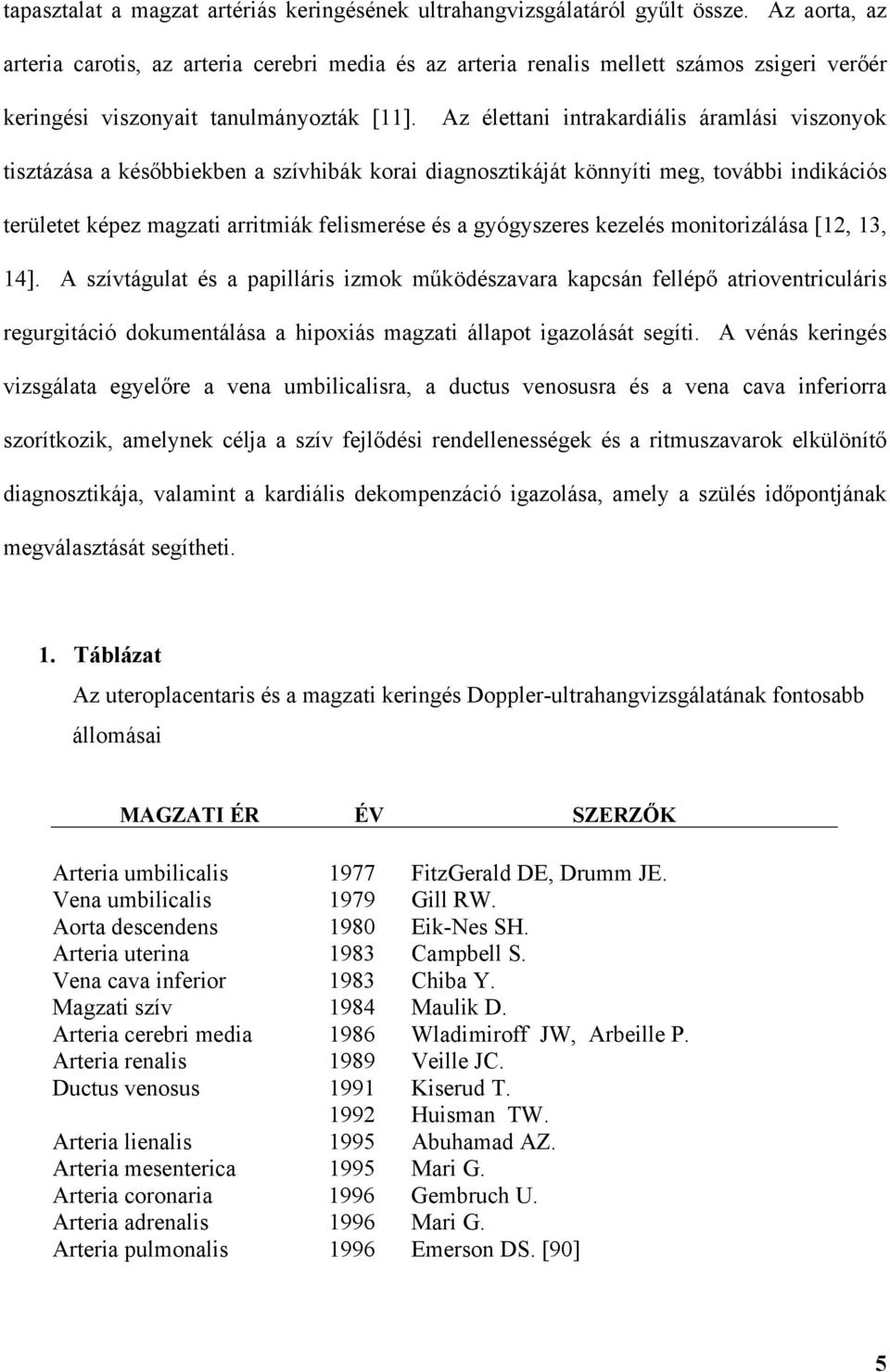
A Doppler-ultrahangvizsgálat szerepe. a kóros lepényi m ködés és a magzati  hipoxia felismerésében - PDF Ingyenes letöltés
A Doppler-ultrahangvizsgálat szerepe. a kóros lepényi m ködés és a magzati hipoxia felismerésében - PDF Ingyenes letöltés
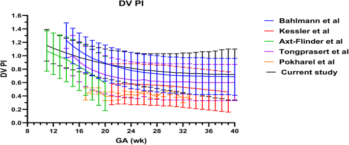
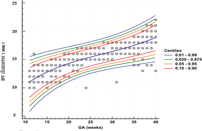
Reference values for ductus venosus flow in normal gestation among an  Egyptian population | Egyptian Journal of Radiology and Nuclear Medicine |  Full Text
Reference values for ductus venosus flow in normal gestation among an Egyptian population | Egyptian Journal of Radiology and Nuclear Medicine | Full Text
RadiologySpirit: How to spot the Normal ductus venosus:

Individual measurements of the ratio S/D for the ductus venosus | Download  Table
Individual measurements of the ratio S/D for the ductus venosus | Download Table

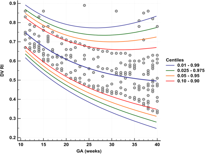
Reference values for ductus venosus flow in normal gestation among an  Egyptian population | Egyptian Journal of Radiology and Nuclear Medicine |  Full Text
Reference values for ductus venosus flow in normal gestation among an Egyptian population | Egyptian Journal of Radiology and Nuclear Medicine | Full Text
Doppler ultrasound - FMF Courses

MAGZATI KERINGÉS ULTRAHANG VIZSGÁLATA by Katalin Tamas - Issuu

MAGZATI KERINGÉS ULTRAHANG VIZSGÁLATA by Katalin Tamas - Issuu

Individual measurements of the ratio (S-A)/TAMX for the ductus venosus |  Download Table
Individual measurements of the ratio (S-A)/TAMX for the ductus venosus | Download Table

Magzati rendellenesség szűrés - magzatom.hu

Reference values for ductus venosus flow in normal gestation among an  Egyptian population | Egyptian Journal of Radiology and Nuclear Medicine |  Full Text
Reference values for ductus venosus flow in normal gestation among an Egyptian population | Egyptian Journal of Radiology and Nuclear Medicine | Full Text

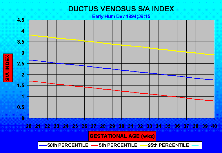
Normal Reference Ranges of Ductus Venosus Doppler Indices in the Period  from 14 to 40 Weeks' Gestation | Semantic Scholar
Normal Reference Ranges of Ductus Venosus Doppler Indices in the Period from 14 to 40 Weeks' Gestation | Semantic Scholar
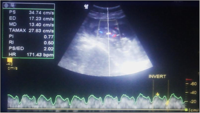
RadiologySpirit: How to spot the Normal ductus venosus:

Reference values for ductus venosus flow in normal gestation among an  Egyptian population | Egyptian Journal of Radiology and Nuclear Medicine |  Full Text
Reference values for ductus venosus flow in normal gestation among an Egyptian population | Egyptian Journal of Radiology and Nuclear Medicine | Full Text
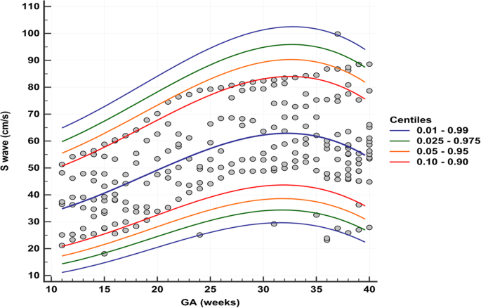

Figure 2 from Fetal ductus venosus pulsatility index and diameter during  20-30 weeks gestation. | Semantic Scholar
Figure 2 from Fetal ductus venosus pulsatility index and diameter during 20-30 weeks gestation. | Semantic Scholar

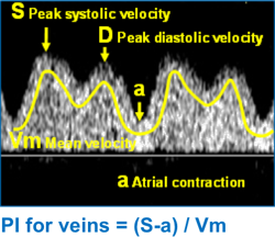
Ductus venosus flow waveform | Radiology Case | Radiopaedia.org